If we hadn’t already mentioned, Julie fits into the high-risk category for two reasons. Because she is over 35 years old, she is in the “advanced maternal age” category – which probably makes a bigger difference in terms of her eggs and the risks for genetic conditions. On top of that, triplets pregnancy is an obvious reason to also put her in the high risk category. The doctor has told her she will most likely fail the glucose test (an indicator of gestational diabetes) and it is certain the babies will be born no later than 36 weeks (40 weeks is full term). With all these risks comes the nice bonus that she will receive great care with close monitoring, which means more frequent ultrasounds.

I don’t know if you’ve ever seen a live fetal ultrasound or not, but to me it is a truly incredible sight. The early ultrasounds at the fertility clinic provide just a taste. Beyond just the chemical test results, pee sticks turning color and your wife missing her period, there’s just something about actually seeing something there. From the ultrasounds we had seen as part of the IUI and IVF process where Julie’s ovaries were being monitored, we had already learned a little bit about how to read the screen. Dark areas are fluid, white areas are tissue. We had a general map in our heads of where the uterus was in relation to the bladder or the ovaries. So at the first ultrasound after getting a positive reading, we knew the three dark blobs were something new.
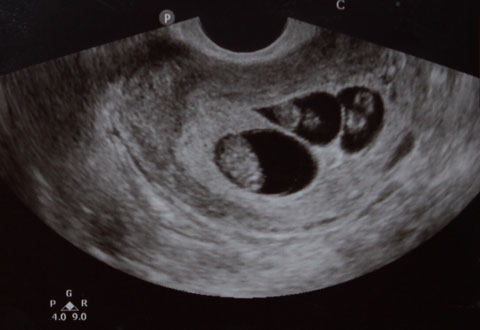
The second ultrasound was particularly cool because it was a new machine and they brought in a new element: sound. For the first time, we not only learned we had triplets, but one at a time we heard each of their heartbeats for baby A, B and C. At about 160 beats per minute, it was music to my ears.
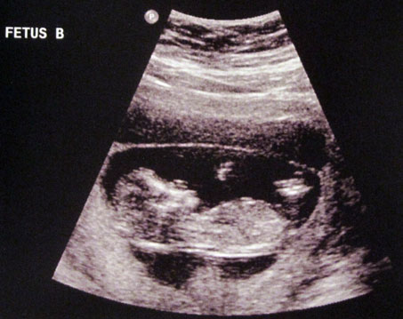
Each ultrasound reveals something new, and we look forward to it each time. At the first ultrasound at the perinatal specialist (11 weeks), we were able to see a lot of activity. Even though it was too early for Julie to feel the babies moving, they were kicking and squirming constantly. We could see their hearts beating more clearly this time.

Baby C always seemed to be a little bigger than Baby A or B, and this time was not different. The nurse referred to Baby C as “the moose” of the bunch. Baby C was enjoying “the penthouse” and had room to grow, while A and B looked like they were competing for space. As the nurse attempted to take measurements of Baby B, Baby A was kicking B in the head. Sibling rivalry begins early.
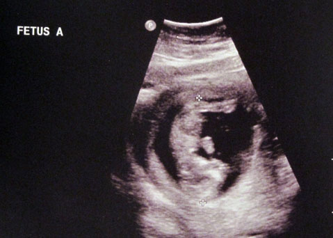
At the next ultrasound, arms and legs were easier to see. Babies were taking a much more “human” appearance, with the shape of the skull and eye sockets becoming more prominent as well. This time, Baby A was doing a headstand for us! I’m starting to think this one will be our little troublemaker.
Because A and B were measuring slightly smaller than Baby C, we asked more about this. The nurse explained that because the babies can be more curled up or stretched out, it is likely they are very close in size or the same size. As she discussed this, she was able to point this out on the screen and it made perfect sense. Before the end of the ultrasound, she also pointed out how organs were becoming visible, and we could even see the tiny dot of a stomach for each baby.
While having triplets means that we will most likely never go through the pregnancy experience again, we are very blessed to have the frequent ultrasounds. I think I enjoy it just as much as Julie, if not more, and I can’t imagine anyone wanting to pass up the opportunity.